images that empower
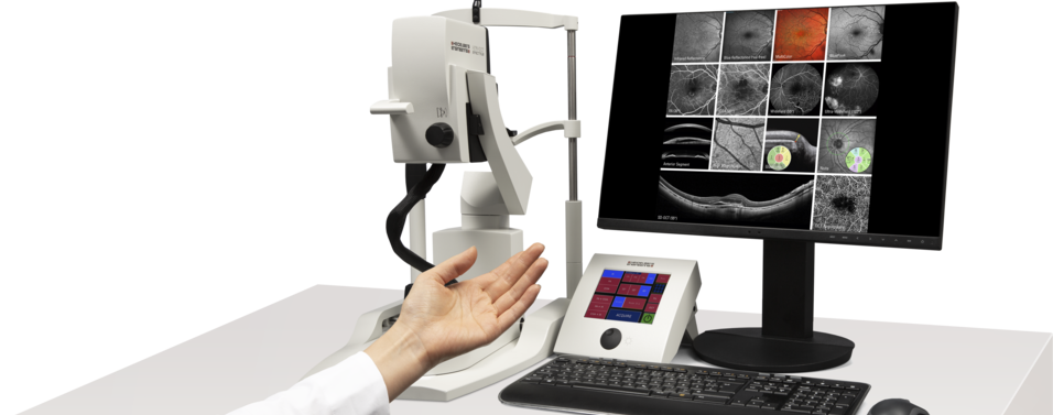
The SPECTRALIS® is an expandable diagnostic imaging platform which combines scanning laser fundus imaging with high-resolution OCT.
Read more ...Glaucoma Module Premium Edition
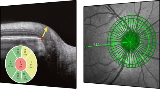
The Glaucoma Module Premium Edition provides a comprehensive and personalized analysis of the optic nerve head, retinal nerve fiber layer, and macular ganglion cell layer by precisely matching unique scan patterns to the fine anatomic structures relevant in glaucoma diagnostics.
Read more ...
MultiColor Module
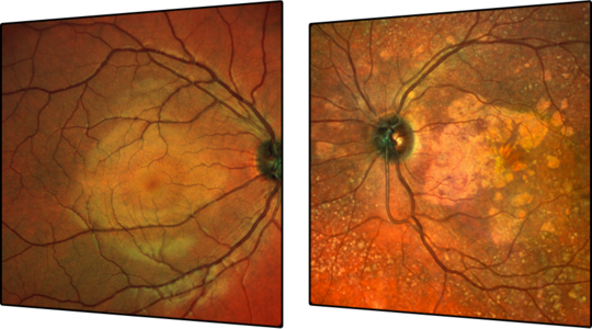
MultiColor is an innovative technology for fundus imaging offering structural detail and clarity not available from traditional fundus photography.
Read more ...
BluePeak Module
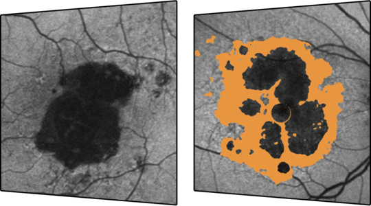
BluePeak is a non-invasive scanning laser fundus imaging modality that provides a map of the retina which can reveal metabolic malfunction of diagnostic significance in many conditions such as AMD.
Read more ...Anterior Segment Module
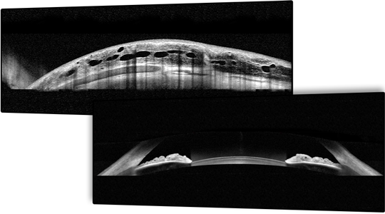
The Anterior Segment Module enables high-resolution OCT imaging of cornea, sclera, and anterior chamber angles.
Read more ...OCT2 Module
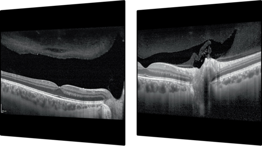
With a scan rate of 85,000 Hz, the OCT2 Module provides more than twice the scanning speed of the SPECTRALIS® .The increased scanning speed supports efficient clinical workflows when performing comprehensive glaucoma evaluations with the Glaucoma Module Premium Edition or high resolution OCT angiography.
Read more ...
OCT Angiography Module
with SHIFT Technology
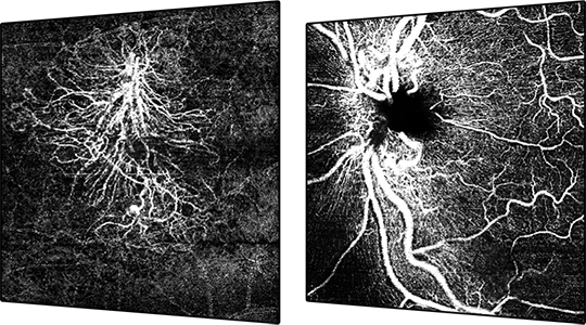
The OCT Angiography Module with SHIFT technology provides a non-invasive imaging technology that delivers a three-dimensional depiction of retinal vascular flow with versatility in field of view, scan speed, and image resolution. With a preset scan speed of 125 kHz, OCTA with SHIFT technology allows you to reduce acquisition time by 50%*.
*Heidelberg Engineering Acquisition Time Comparison Video: SPECTRALIS with OCT2 Module at 85 kHz vs. SPECTRALIS with SHIFT Module at 125 kHz.
Read more ...Scanning Laser Angiography
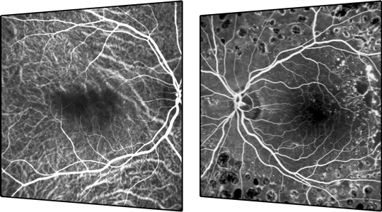
SPECTRALIS® scanning laser angiography provides high-resolution diagnostic images and video sequences showing the dynamic movement of dye through the vessels and minute details of the parafoveal capillary network.
Read more ...Widefield Imaging Module
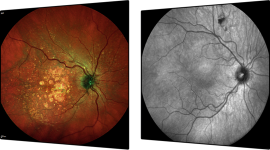
The Widefield Imaging Module provides the standard field of view of a mydriatic fundus camera for all SPECTRALIS® fundus imaging modalities, simplifying diagnostic protocols and facilitating detection of peripheral pathology.
Read more ...Ultra-Widefield Imaging Module
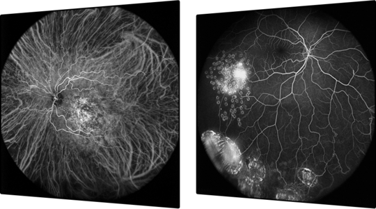
The Ultra-Widefield Imaging Module delivers evenly illuminated, high-contrast scanning laser angiography images from the macula through the periphery.
Read more ...High Magnification Module
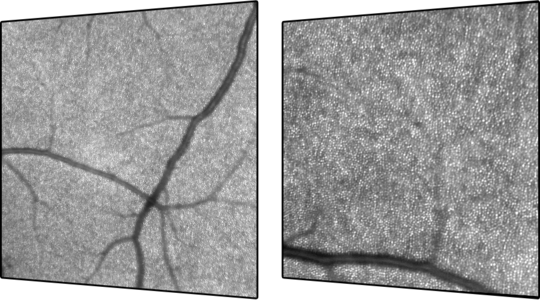
The High Magnification Module enables non-invasive, high-resolution cSLO imaging and visualization of previously imperceptible retinal microstructures.
Read more ...