The important role of advanced imaging in identifying ocular indications of systemic conditions
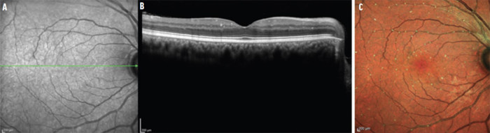
Image courtesy: Robert Sergott, MD
In this article, Dr. Robert Sergott highlights the value of SPECTRALIS multicolor OCT imaging in the diagnosis and management of patients with systemic disease. Based on his extensive experience in neuro-ophthalmology, he provides tips and examples for applying this advanced imaging in the diagnosis of emboli and vaso-occlusive disease, neuromyelitis optica spectrum disorder, and dermatomyositis.
For more valuable information about our products, educational offerings, learning materials and events
Subscribe to our Newsletter orContact us