Andrew Tatham
“Spectralis allows me to improve my ability to diagnose glaucoma”
How are glaucoma patients referred into the hospital?
Our hospital glaucoma team consists of ophthalmologists, optometrists,ophthalmic technicians and nurses. We run very busy clinics due to the high prevalence of glaucoma and due to the service being the sole provider of glaucoma care for a population of close to a million. Patients with suspected glaucoma are referred to the glaucoma clinic by community optometrists using an electronic referral system, with a requirement for referrals to include IOP measurements from applanation tonometry, disc photographs, and copies of visual fields. This helps reduce false positive referrals and ensures appointments for patients at higher risk of visual loss can be prioritised. Many referrals also include information from OCT.
“When viewing an OCT, it is essential to look at the whole scan, not just the OCT print-out or PDF report. We need to see the big picture”
How do you use OCT in the diagnosis and management of glaucoma?
When diagnosing glaucoma, my approach is to consider the clinical information, examination findings and visual fields first. I try to determine the probability that a patient has glaucoma and then review the results of the OCT to see if it agrees with my suspicions. In my opinion, when viewing an OCT, it is essential to look at the whole scan, not just the OCT print-out or PDF report. We need to see the big picture. Only by examining the whole scan can subtle and localised changes be identified.When we examine the results of a visual field test, glaucoma specialists do not rely solely on summary measures such as mean deviation. Doing so would miss localised areas of abnormality and reduce our ability to detect progression. It should be the same with OCT and I find it best to have it networked to my office so that I can look at more than the average retinal nerve fiber layer (RNFL) thickness and see the images themselves.Reviewing all of the available data,including rim width, RNFL and macular thickness is helpful, especially in cases of diagnostic uncertainty.
“Spectralis supplies the highest quality and most reproducible images”
Which OCT platform do you use?
We use the Spectralis with Glaucoma Module Premium Edition for all patients with glaucoma and suspected glaucoma and it has become an essential tool in our clinic. My experience is that the Spectralis allows me to improve my ability to diagnose glaucoma, particularly to identify early glaucoma or glaucoma in patients who struggle with visual field testing. OCT also helps to improve the confidence with which one can discharge patients back to the care of the optometrist when all is normal. I also use the Spectralis to detect glaucoma progression. In cases of diagnostic uncertainty, looking for change over time can be a very useful way to confirm the diagnosis and inpatients with confirmed glaucoma, faster rates of change on OCT have been shown to be a risk factor of progressive visual loss. An important point is that when trying to detect change, we need to be confident that what we are seeing is genuine change,as opposed to visit to visit variability,for example, due to variation in the placement of the OCT scan. The AutoRescan feature of the Spectralis gives me confidence on whether I am seeing real progression or not.
What do you like about SPECTRALIS?
I have been fortunate to try almost every OCT platform, but I believe the Spectralis supplies the highest quality and most reproducible images. It enables us to image all the structures relevant in glaucoma on one platform –RNFL, ganglion cell layer, full retinal thickness and neuroretinal rim. We now know that imaging the macular can reveal glaucomatous changes that may be missed with RNFL imaging alone. The multimodal approach offered by Spectralis enables me to look at agreement between scans to confirm a diagnosis.
Spectralis provides me with large images of the wholescan and the image is so clear that it’s possible to identify glaucomatous changes with the naked eye. Spectralis also allows me to check the segmentation, which we do as part of a mandatory routine because there is a chance the segmentation has been confounded by, for example, a prominent posterior hyaloid. It doesn’t happen often, but when it does it is nice that Spectralis offers the option to see it and manually adjust the segmentation. Heidelberg Engineering is not always the first to market, but they really take their time to ensure their products are right and clinically relevant when they are released. They’re at the forefront of innovation – using Bruch’s Membrane Opening as a reference point was a big leap forward for example– and the support and training that has been offered has been extremely good.
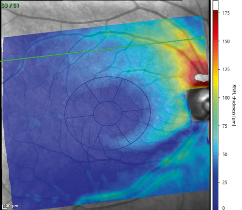
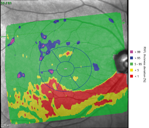
Thickness map (left) and deviation map (right) showing arcuate temporal inferior RNFL thinning in a glaucoma patient.
More information
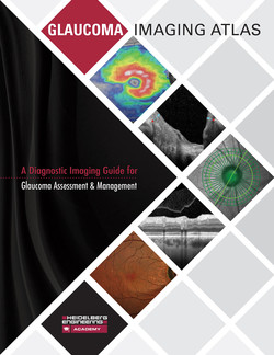
If you are interested to learn more about how OCT imaging can be used as part of the assessment in glaucoma, you may be interested to purchase the Glaucoma Imaging Atlas, which is a comprehensive diagnostic guide for glaucoma assessment and management featuring 29 contributors from five countries and 29 detailed patient case studies. Purchase the Atlas online at www.glaucoma-imaging-atlas.com
Andrew Tatham, Consultant Ophthalmic Surgeon
Princess Alexandra Eye Pavilion
Edinburgh
Advertorial written and published by Optometry Today magazine.