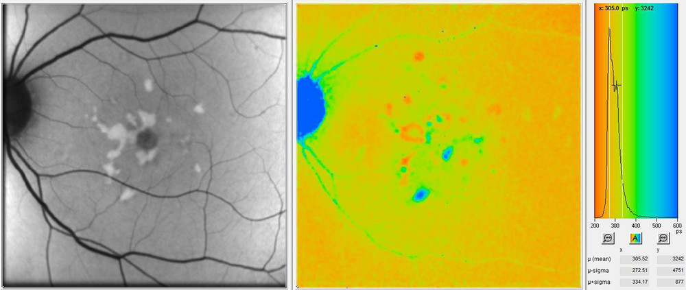
Novel fluorescence lifetime imaging may complement BluePeak for prediction of progression in Stargardt disease
Since its introduction, BluePeak blue laser autofluorescence has become a widely established scanning laser fundus imaging modality to provide a map of the retina which can reveal metabolic malfunction of diagnostic significance in many conditions. Diagnosing and monitoring early AMD and late stages of geographic atrophy as well as hereditary diseases such as Stargardt disease are just a few examples of the diagnostic value of this non-invasive imaging modality.

However, whereas BluePeak provides a two-dimensional map of the intensity of the autofluorescence signal, a novel imaging modality called “fluorescence lifetime imaging ophthalmoscopy” (FLIO)* measures the time-dependency of fundus autofluorescence emission. To illustrate the difference, imagine examining a firefly with both imaging modalities; BluePeak would show how strongly it glows and FLIO would show how long it glows over a period of time. An example of how both imaging modalities could complement each other for diagnostic purposes is shown in the figure above.
In a recent publication1, a study group in Switzerland examined autofluorescence lifetime characteristics in Stargardt disease to investigate potential prognostic markers for disease activity and progression. They demonstrated that certain changes in fluorescence lifetimes were predictive of disease progression. These changes were not seen on standard BluePeak images. The authors conclude that changes seen on FLIO images over time may provide a useful tool to predict disease progression and may be used to assess therapeutic effects in upcoming clinical trials for those patient groups.