Subbasal nerve plexus: Examine, explore, analyze with HRT3 RCM
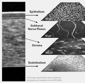
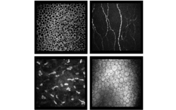
 HRT3 RCM is a confocal in vivo corneal microscope that allows you to acquire unique en face images of the corneal layers. This gives you insights into cell structures and potential pathological changes within the entire cornea.
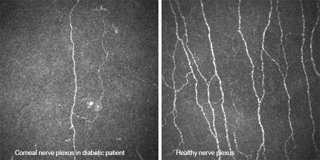
In patients with diabetes mellitus, damage to the peripheral nervous system can occur. This diabetic neuropathy can lead to functional disorders. Indications of these changes may be visible in the subbasal nerve plexus. HRT3 RCM supports you in the in vivo examination of the subbasal nerve plexus. The following images show the difference between the corneal nerve structure of a diabetes patient respective to a healthy eye.

Learn more about further clinical applications of HRT3 RCM in our upcoming newsletters, which will feature clinical cases from eye care specialists around the world. In addition, from 2-4 October 2020, you can find out more about our solutions for anterior segment diagnostics at our virtual exhibition stands at EURETINA or ESCRS. Both events will take place online this year. We are looking forward to meeting you!
 HRT3 RCM - In vivo corneal microscope
Learn more about HRT3 RCM and explore the impressive high-resolution images in the image gallery.